Oesophagostomum columbianum (nodule worm) is a gastrointestinal nematode that inhabits the small and large intestine of ruminants. The nodule worm is found predominately throughout summer rainfall areas of Australia and is considered the most pathogenic or disease forming species of Oesophagostomum. Nodule worm is not particularly common and is typically seen during January in mixed infections (generally less than 10% of the worm burden). However, it can cause severe pathological changes to the intestine that results in impaired intestinal absorption. Infections, especially in young stock will exhibit clinical signs of diarrhoea, depressed appetite and abdominal pain with significant weight loss over time.
In the laboratory, diagnosis of infection is performed by conducting worm egg counts and culturing faeces for incubation at 25–27°C for 7 days, allowing eggs to develop to infective third stage larvae (L3). It is the L3 larvae that are stained, allowing identification of worm species (and differentiation from the less pathogenic O. venulosum) under high magnification by trained laboratory personnel.
Unlike most worm species however, it takes approximately 40 days after ingestion of the L3 larvae to detect eggs in the faeces. This is the time required for larvae to develop to an adult worm. By this stage, noticeable clinical effects have already resulted. Performing routine worm egg count monitoring and being aware of worm species in your flock can greatly assist with appropriate management of such worms.
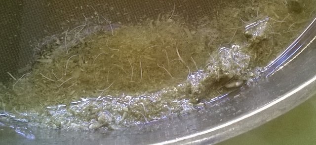
Performing further investigations at the post mortem level gives insight into the extent of damage created by this worm species. As nodule worm resides predominately in (but not limited to) the large intestine (LI), examination of the LI contents will reveal the adult worms. Unlike most worms, the adult worms are clearly visible with the naked eye (Figure 1). Sifting through the contents of the large intestine and counting adult worms gives a definitive indication of worm burden.

When the small and large intestine are washed of contents and the tissue is examined more closely, the gross pathology attributed to infection can be seen. Intestinal lesions described as green pus-filled nodules are evident due to the animal’s immune system attempting to combat the infection. These nodules can extend the entire length of the intestine, impairing normal organ function. When an animal grazes infected pastures, the ingested infective larvae (L3) migrate to the small intestine then subsequently enter the intestinal wall. Approximately 5–9 days post-infection, most larvae re-enter the small intestine and migrate further to the large intestine and either develop to an adult worm or, once again, penetrate and enter the large intestinal wall. The larvae may reside in the wall for some time (weeks or months). The intensity of the immune system and resulting pathology (nodules) has been noted to increase in those animals that have had prior infections. As seen in Figures 2 and 3, the green pus-filled nodules (some are highlighted in the white circles on the images) are clearly seen in both the small and large intestine along with red inflamed tissue.
Such an immune reaction is evident 1–2 weeks after ingestion of larvae with infected animals displaying clinical signs of intermittent diarrhoea and loss of appetite. In the meat industry, it is the green pus-filled nodules that make the intestine unsuitable for sausage casing at slaughter.
Looking at the adult worm under high magnification assists with identification of species. In Figures 4 and 5, the characteristic anterior (head) and posterior (tail) ends of this male adult worm can be viewed.
Although the number of reported cases are low, the pathogenicity of this worm species in young sheep is evident. Furthermore, infections display similar signs to the black scour worm and hence this worm is often overlooked. Regular worm egg count monitoring along with nutritional and pasture management can assist in reducing the risk of infection.